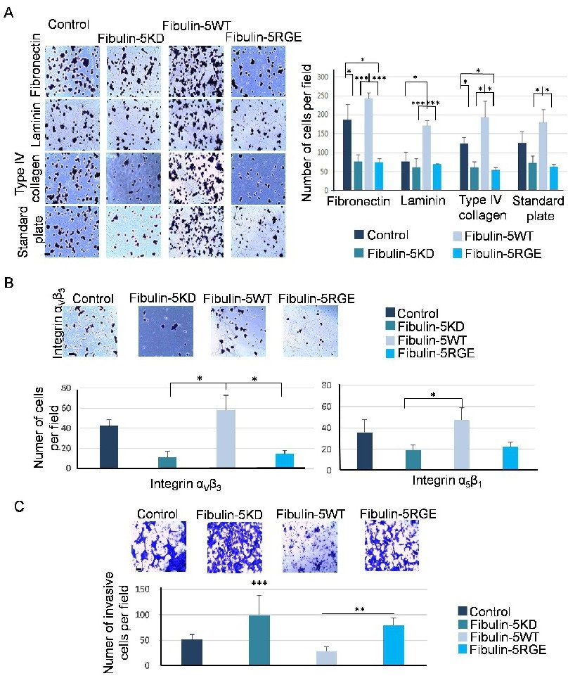
Fig. 4. RGD-to-RGE change in fibulin-5 alters the adhesion profile and the invasive ability of 4T1 cells. A. Left, representative images showing attachment of the indicated 4T1 cells to the ECM components fibronectin, laminin and type IV collagen. Adhesion to standard culture plates was also examined. Bar, 10 µm. Right, graphical representation of the number of attached cells. Exogenous expression of an intact fibulin-5 (Fibulin-5WT) increases the number of attached 4T1 cells in all assayed conditions. B. Ability of the 4T1 cells used in this work to bind to ααvβ3 integrin was also examined. Top, representative images of the indicated 4T1 cells attached to the αvβ3 integrin. Bar, 10 µm. Bottom, graphical representation of the number of attached cells to the αvβ3 and α5β1 integrins as indicated. Exogenous expression of an intact fibulin-5 (Fibulin-5WT) also increases adhesion of 4T1 cells (color code as in C) to the αvβ3 integrin. C. Top, representative pictures showing invasive 4T1 cells as indicated using Matrigel-coated invasion chambers. Bar, 10 µm. Bottom, quantification of the number of invasive cells.